
KPV: Inflammatory Signaling, Barrier Tissues, and Key Findings
Research Area: Immune & Inflammatory Signaling Research
KPV is a three-amino-acid peptide made of lysine, proline, and valine. It matches the final three amino acids of alpha-melanocyte-stimulating hormone, usually shortened to alpha-MSH. KPV is studied because this very short sequence retains much of the parent hormone’s inflammatory-signaling activity without carrying the sequence associated with pigment production.
The useful research question is not whether KPV broadly “stops inflammation.” It is which signaling pathways change, which cells respond, and whether molecular changes are accompanied by preserved barrier structure and normal tissue behavior.
Alpha-MSH connects nervous, pigment, and immune signaling
Alpha-MSH is produced from a larger precursor protein and can signal through melanocortin receptors. These receptors appear in several tissues, including immune cells and barrier tissues. Alpha-MSH-related signaling can influence pigmentation, inflammation, cell movement, oxidative stress, and cytokine production.
KPV is unusual because it lacks the full sequence normally required for several known melanocortin-receptor interactions yet retains inflammatory effects in published research. That has led researchers to study both receptor-dependent and receptor-independent explanations.
NF-κB is a central measurement
NF-κB is a family of proteins that helps turn inflammatory genes on or off. When certain receptors detect stress signals, NF-κB can move into the nucleus and influence cytokines, adhesion molecules, survival proteins, and other parts of the response.
KPV and related alpha-MSH research often examines whether NF-κB activation changes. A complete interpretation may include nuclear movement, DNA binding, target-gene activity, and the downstream cytokines or cell behavior produced by that signal.
Barrier tissues do more than separate spaces
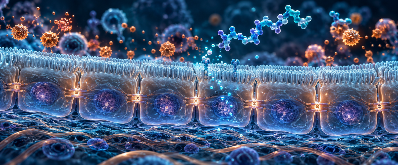
Epithelial cells form continuous layers at the skin, airway, and intestinal lining. They are physical barriers, but they also sense their environment, release cytokines, and communicate with immune cells. Neighboring epithelial cells are connected by structures called tight junctions.
Inflammatory signals can change tight-junction proteins and make a barrier more permeable, meaning material crosses the cell layer more easily. Researchers therefore pair molecular measurements with physical tests such as electrical resistance across the layer or movement of tracer molecules.
Cytokines and cell movement help define the response
Cytokines carry instructions between cells. Chemokines are signals that guide cell movement. Adhesion molecules help circulating immune cells slow down, attach, and enter tissue. Alpha-MSH-related pathways can affect each part of this traffic system.
A lower cytokine value is only one piece of evidence. Researchers also ask whether the right cells reached the tissue, whether disrupted material was removed, whether the barrier remained intact, and whether the response moved toward resolution.
KPV and the resolution side of inflammation
Resolution is the active transition from an inflammatory response toward cleanup and restored tissue balance. It includes stopping further cell recruitment, removing spent immune cells, clearing debris, and supporting repair. This is different from simply blocking the first signal.
KPV research is useful when it connects pathway measurements such as NF-κB and cytokines with barrier function and cell behavior. That combination helps show whether signaling changed in a way that matters to the tissue model being studied.
Key findings to understand
- KPV is the C-terminal tripeptide of alpha-MSH. C-terminal means the end of the peptide chain.
- The short sequence retains inflammatory-signaling activity. This allows researchers to separate those effects from the parent hormone’s pigment-related biology.
- NF-κB is a repeated mechanistic focus. Changes in NF-κB can influence cytokines, adhesion molecules, and other inflammatory genes.
- Barrier function requires physical measurements. Tight-junction proteins, electrical resistance, and permeability show whether a cell layer changed structurally.
- Inflammatory signaling and antimicrobial activity are separate questions. Each requires its own controls and measurements.
Measurements that answer different questions
| Measurement | What it helps answer |
|---|---|
| NF-κB nuclear activity | Whether a major inflammatory gene-control pathway changed |
| Cytokines and chemokines | Which instructions and cell-recruitment signals were released |
| Cell migration | Whether immune or tissue cells changed their movement |
| Tight-junction proteins | Whether the structures sealing neighboring barrier cells changed |
| Electrical resistance | Whether the barrier became easier or harder for ions to cross |
| Permeability tracers | Whether selected molecules crossed the barrier layer |
How KPV differs from Thymosin Alpha-1 and ARA-290
KPV is centered on inflammatory signaling and barrier tissues. Thymosin Alpha-1 is centered on coordination among dendritic cells, pattern-recognition pathways, and T cells. ARA-290 is centered on the innate repair receptor in stressed tissue.
See Thymosin Alpha-1 vs. KPV vs. ARA-290: Immune Research Compared, or return to Immune & Inflammatory Signaling Research.
Common questions
Is KPV the same as alpha-MSH?
No. KPV is the final three-amino-acid fragment of the longer alpha-MSH peptide. It preserves selected research properties without representing every function of the full hormone.
Does lower NF-κB activity prove that a barrier recovered?
No. NF-κB describes signaling. Barrier recovery requires structural and functional measurements such as tight-junction organization and permeability.
Why study skin, airway, and intestinal barriers separately?
They share epithelial features but differ in cell types, exposures, local immune populations, and normal permeability. A finding in one barrier should not automatically be assigned to another.
Research product
View the CellTek Labs KPV research product.
References
- Alpha-MSH-related tripeptides and inflammatory signaling
- Inflammatory effects of alpha-MSH-related peptides and KPV
- Dissection of alpha-MSH and KPV inflammatory signaling
- KPV and intestinal inflammatory signaling
- KPV and airway epithelial signaling
Research and educational content only. CellTek products are not for human or veterinary use.
