
Thymosin Alpha-1 vs. KPV vs. ARA-290: Immune Research Compared
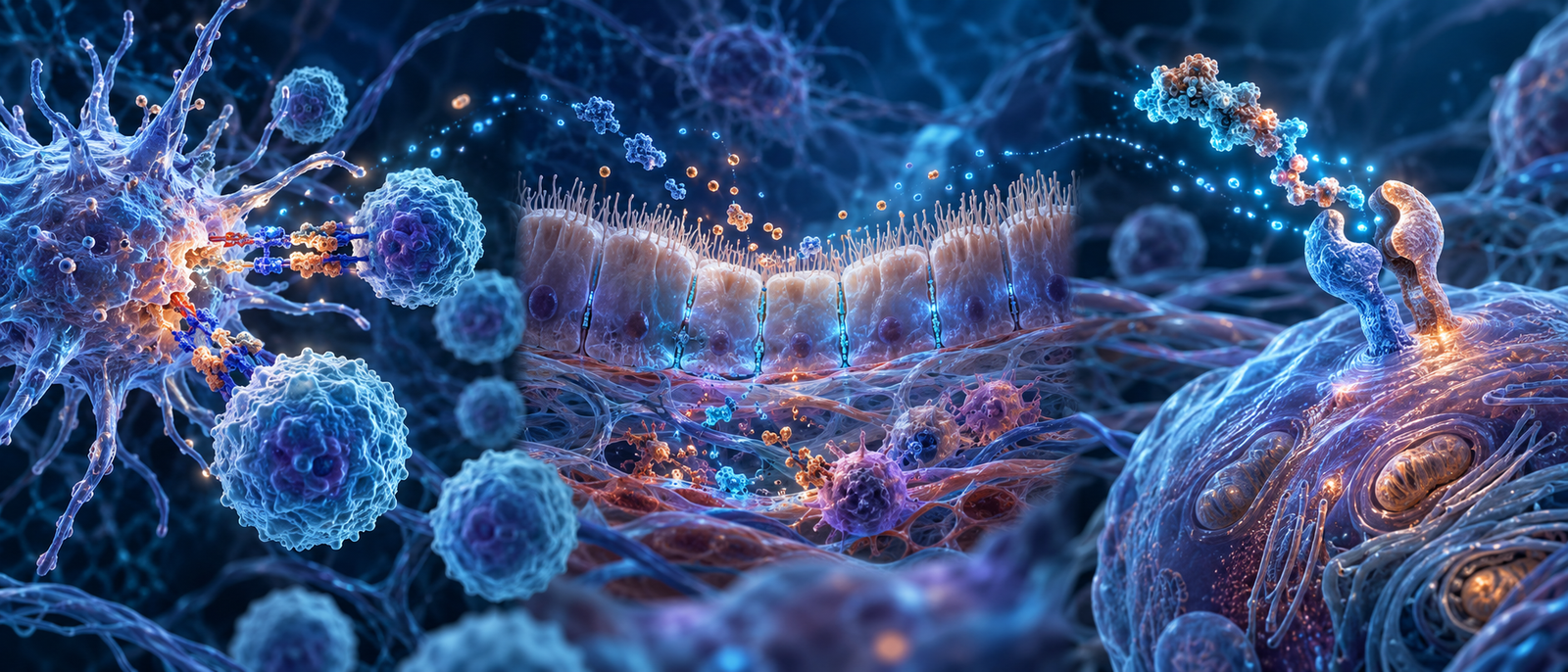
Research Area: Immune & Inflammatory Signaling Research
Thymosin Alpha-1, KPV, and ARA-290 all appear in immune or inflammatory research, but they begin at different points in the system. Thymosin Alpha-1 is studied for immune-cell coordination. KPV is studied for inflammatory gene signaling and barrier tissues. ARA-290 is studied for a tissue-protective receptor that appears during cellular stress.
Calling all three “anti-inflammatory” removes the information needed to design a useful comparison. The better question is whether the study concerns communication among immune cells, control of a barrier response, or stress-linked protection and repair.
The short comparison
| Thymosin Alpha-1 | KPV | ARA-290 | |
|---|---|---|---|
| What it is | A 28-amino-acid thymic peptide | A three-amino-acid fragment related to alpha-MSH | An 11-amino-acid peptide designed from erythropoietin’s helix B surface |
| Main question | How does innate recognition shape adaptive immune coordination? | How do inflammatory pathways and barrier tissues respond? | How does the innate repair receptor signal after tissue stress? |
| Primary system | Dendritic cells, TLRs, T cells, and tolerance | NF-κB, cytokines, migration, and epithelial barriers | EPOR/CD131 signaling, protection, and repair |
| Essential control | Measure function as well as immune markers | Measure barrier function as well as cytokines | Separate the repair receptor from red-blood-cell signaling |
Thymosin Alpha-1 starts with immune coordination
Thymosin Alpha-1 research emphasizes the bridge between innate and adaptive immunity. Dendritic cells receive early signals through pattern-recognition systems such as Toll-like receptors, then influence how T cells respond.
The most useful measurements follow that chain: receptor signaling, dendritic-cell state, cytokine pattern, T-cell subsets, and immune-cell function. This approach distinguishes coordinated regulation from a nonspecific increase in activity.
KPV starts with inflammatory and barrier signaling
KPV is the C-terminal three-amino-acid sequence of alpha-MSH. Its research commonly examines NF-κB, cytokines, adhesion signals, cell movement, and the behavior of epithelial barriers.
A KPV-centered comparison should not stop at molecular markers. Tight-junction proteins, electrical resistance, or permeability tests show whether a change in signaling was accompanied by a change in the physical cell layer.
ARA-290 starts with a stress-linked receptor
ARA-290 is designed to interact with the innate repair receptor, a pair made from EPOR and CD131 receptor subunits. This receptor is studied in stressed tissue and is different from the paired EPOR receptor associated with red-blood-cell production.
Useful measurements establish receptor involvement, downstream signaling, inflammatory markers, and a tissue-specific structural or functional outcome. Controls should also verify that the intended pathway remains separate from erythropoiesis.
Where the pathways can overlap
All three pathways can change cytokines and other inflammatory markers. That overlap does not make the mechanisms identical. Thymosin Alpha-1 may change how immune cells interpret and pass information. KPV may change NF-κB-related signaling at a barrier. ARA-290 may change how stressed tissue responds through a specific paired receptor.
The overlap becomes useful when researchers follow cause and sequence. A receptor signal may change gene activity, which changes cytokines, which changes cell movement, which changes tissue structure. Measuring only the final cytokine value hides that pathway.
Which comparison fits the question?
- Choose Thymosin Alpha-1-centered measurements for dendritic cells, Toll-like receptor pathways, T-cell coordination, interferons, or immune tolerance.
- Choose KPV-centered measurements for NF-κB, cytokines, epithelial tight junctions, permeability, or inflammatory cell migration.
- Choose ARA-290-centered measurements for EPOR/CD131 signaling, cellular stress, programmed cell death, tissue protection, or later repair.
- Combine pathways only with a clear sequence. State which signal comes first, which intermediate steps are measured, and what tissue-level outcome would support the proposed connection.
Key distinctions
| Question | Best starting point | Why |
|---|---|---|
| Did innate recognition change later T-cell behavior? | Thymosin Alpha-1 | Its research connects dendritic-cell and TLR signaling with adaptive responses. |
| Did inflammatory signaling alter a barrier? | KPV | Its research links NF-κB and cytokines with epithelial tissues. |
| Did stressed tissue activate a protection-and-repair pathway? | ARA-290 | It is designed for the EPOR/CD131 innate repair receptor. |
| Did inflammation simply become “good” or “bad”? | None alone | Inflammation changes across time, location, cell type, and resolution stage. |
Common questions
Do these compounds use the same receptor?
No. Their research begins with different signaling systems. Shared downstream markers such as NF-κB or cytokines do not mean the initiating receptor is the same.
Which one is an immune “booster”?
That label is too broad to be useful. Immune systems need recognition, activation, restraint, tolerance, resolution, and repair. Each compound should be studied with measurements matched to its proposed pathway.
Can the same cytokine be used to compare all three?
It can provide one shared measurement, but it does not identify the mechanism. Receptor activity, cell identity, timing, and tissue function are needed to explain why the cytokine changed.
Research products
Return to Immune & Inflammatory Signaling Research for the complete pathway overview.
References
- Innate and adaptive immunity interplay
- Immune modulation with Thymosin Alpha-1
- Alpha-MSH-related tripeptides and inflammatory signaling
- ARA-290 and the innate repair receptor
- Resolution of inflammation
Research and educational content only. CellTek products are not for human or veterinary use.
