
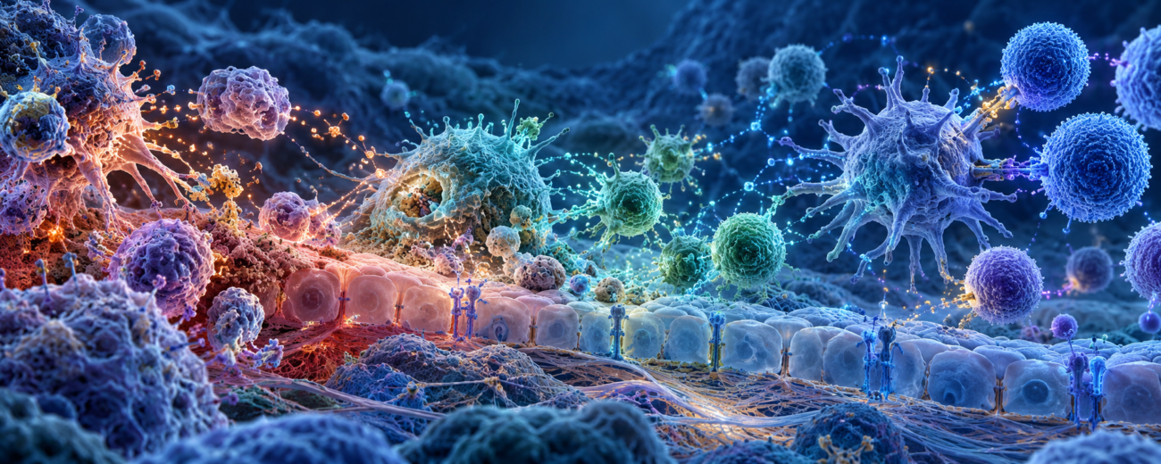
Immune & Inflammatory Signaling Research
The immune system is a communication network. Cells recognize possible damage, send chemical instructions, recruit help, remove disrupted material, and then coordinate a return toward normal tissue function. Inflammation is one part of that sequence—not a separate enemy and not a simple dial where less is always better.
This research area separates three different questions. Thymosin Alpha-1 is studied for coordination between innate and adaptive immunity. KPV is studied for inflammatory signaling at barrier tissues. ARA-290 is studied for activation of a tissue-protective receptor that appears after stress or injury.
Inflammation has a beginning, a working phase, and a resolution phase
An inflammatory response begins when cells detect patterns associated with damage or possible threats. Blood vessels and nearby tissues change their behavior. Immune cells move toward the affected area. Chemical signals help determine which cells arrive, what they do, and how long they remain.
Ending the response is not simply the absence of activation. Resolution is an active program. Incoming cells stop accumulating, spent cells and debris are removed, signaling changes, and tissue repair becomes the priority. A response that starts too weakly may fail to contain a problem; one that remains active too long may disturb healthy tissue.
Innate and adaptive immunity answer different questions
The innate immune system responds quickly to broad patterns. It includes barrier cells, macrophages, neutrophils, dendritic cells, natural killer cells, and molecular systems that recognize common signs of stress. The adaptive immune system develops highly specific responses through T cells and B cells and can retain immune memory.
These branches constantly exchange information. Dendritic cells are an important bridge. They sample their surroundings, process material, and present selected pieces to T cells. The result depends on the surrounding signals: activation, restraint, tolerance, and memory all require context.
Receptors turn outside information into cellular instructions
A receptor is a protein that recognizes a particular type of signal and starts activity inside the cell. Toll-like receptors, usually shortened to TLRs, recognize broad molecular patterns and help initiate innate responses. Other receptors respond to cytokines, growth signals, or tissue stress.
Once a receptor is activated, the message moves through a signaling pathway. Proteins switch one another on or off until the signal reaches the nucleus, where gene activity may change. Measuring a receptor without measuring the downstream pathway can miss whether the message was actually carried through the cell.
Cytokines and chemokines are instructions, not simple good-or-bad markers
Cytokines are small proteins that cells use to change the behavior of other cells. Chemokines are a related group that helps guide cell movement. A cytokine can increase activity in one setting, limit it in another, or produce different outcomes in different cell types.
This is why a lower cytokine measurement does not automatically prove that a system improved. Researchers consider which cytokine changed, where it was measured, which cells produced it, what happened earlier, and whether the tissue successfully moved into resolution.
NF-κB coordinates many inflammatory genes
NF-κB is a family of proteins that helps control genes involved in inflammation, cell survival, and immune activity. In a resting cell, NF-κB is often held outside the nucleus. Receptor signals can release it, allowing it to enter the nucleus and change gene activity.
NF-κB is useful because it links many receptors with cytokine production and other responses. It is not an on-off definition of inflammation. Researchers may measure NF-κB movement into the nucleus, DNA binding, target-gene activity, cytokines, and the behavior of the tissue barrier together.
Barrier tissues are active immune participants
Skin, airway surfaces, and the intestinal lining do more than form walls. Their cells recognize signals, produce cytokines, communicate with immune cells, and regulate what can pass between neighboring cells. The protein structures that seal those spaces are called tight junctions.
Barrier research may measure electrical resistance across a cell layer, movement of tracer molecules, tight-junction proteins, NF-κB activity, and cytokine release. These measurements distinguish a signaling change from a physical change in barrier function.
Three compounds, three starting mechanisms
Thymosin Alpha-1 is a 28-amino-acid peptide connected with dendritic cells, Toll-like receptor pathways, T-cell activity, and immune tolerance. Its research focus is coordination: how innate recognition shapes a later adaptive response without reducing immunity to a simple “boost.”
KPV is the three-amino-acid sequence lysine-proline-valine and is related to the end of alpha-melanocyte-stimulating hormone. It retains inflammatory-signaling activity without the hormone’s pigment-related sequence. Research often examines NF-κB, cytokines, cell migration, and barrier tissues.
ARA-290, also known as cibinetide, is an 11-amino-acid peptide designed from part of erythropoietin’s three-dimensional structure. It is studied for selective signaling through the innate repair receptor, a paired receptor that appears in stressed tissue. This pathway is distinct from erythropoietin’s red-blood-cell-producing receptor.
How the research questions differ
| Compound | Main research focus | Useful measurements |
|---|---|---|
| Thymosin Alpha-1 | Coordination between innate recognition and adaptive responses | Dendritic-cell activity, TLR signaling, T-cell subsets, interferons, cytokines, and tolerance-related pathways |
| KPV | Inflammatory signaling and barrier-tissue responses | NF-κB activity, cytokines, cell migration, tight-junction proteins, permeability, and oxidative-stress markers |
| ARA-290 | Innate repair receptor signaling after cellular stress | EPOR/CD131 receptor activity, inflammatory pathways, apoptosis signals, tissue integrity, and repair markers |
Key findings researchers examine
- Immune modulation is not the same as immune stimulation. A compound may change how cells coordinate, differentiate, or tolerate signals rather than simply increasing activity.
- Thymosin Alpha-1 connects recognition with response. Research emphasizes dendritic-cell and Toll-like receptor pathways that can shape later T-cell activity.
- KPV retains inflammatory-signaling activity in a very short sequence. Its research connects alpha-MSH-related biology with NF-κB, cytokines, cell movement, and barrier tissues.
- ARA-290 separates tissue-protective signaling from red-blood-cell production. Its research centers on the EPOR/CD131 innate repair receptor rather than the receptor pair responsible for producing red blood cells.
- Resolution is an active outcome. Removing debris, changing cell traffic, and rebuilding tissue require signals of their own.
Explore the immune and inflammatory research articles
- Thymosin Alpha-1: Immune Coordination and Key Findings — How dendritic cells, Toll-like receptors, T cells, and immune tolerance fit together.
- KPV: Inflammatory Signaling, Barrier Tissues, and Key Findings — A guide to alpha-MSH-related signaling, NF-κB, cytokines, and barrier measurements.
- ARA-290: The Innate Repair Receptor and Tissue-Protective Signaling — Why this peptide is studied separately from erythropoietin’s blood-cell pathway.
- Thymosin Alpha-1 vs. KPV vs. ARA-290: Immune Research Compared — A side-by-side guide to immune coordination, barrier signaling, and tissue-protective responses.
Choosing a useful research comparison
For immune-cell coordination, begin with Thymosin Alpha-1 and measurements that connect dendritic-cell signaling to T-cell outcomes. For barrier-centered inflammation, begin with KPV and pair molecular markers with physical barrier measurements. For stress-linked tissue protection, begin with ARA-290 and verify both receptor activity and the downstream state of the tissue.
A single cytokine should rarely stand alone. A stronger design combines a pathway marker, a cell-behavior measurement, and a tissue-level result. Timing also matters: the same signal may be useful early, unnecessary later, or disruptive if it persists.
Common questions
Is inflammation always harmful?
No. It helps contain damage, recruit immune cells, and begin repair. The research question is whether the response is appropriate in strength, location, duration, and resolution.
What is immune tolerance?
Immune tolerance is controlled restraint toward the body’s own material and other signals that should not trigger a damaging response. It is an active part of immune regulation, not a failure to respond.
Does a lower cytokine level prove that tissue recovered?
No. Cytokines describe communication. Tissue recovery also requires restored structure, normal barrier function, appropriate cell populations, and successful resolution.
Do Thymosin Alpha-1, KPV, and ARA-290 use the same receptor?
No. Thymosin Alpha-1 research involves several immune-signaling systems, including Toll-like receptor pathways. KPV is studied through alpha-MSH-related inflammatory pathways. ARA-290 is designed to signal through the EPOR/CD131 innate repair receptor.
Learn More by Product
Choose a product to view its catalog page, then open the related article for a focused explanation of its pathway, key findings, and comparisons.
Compare the group: Compare Thymosin Alpha-1, KPV, and ARA-290.
References
- Resolution of inflammation as an organizing principle
- Innate and adaptive immunity interplay
- Immune modulation with Thymosin Alpha-1
- Thymosin Alpha-1, inflammation, immunity, and tolerance
- Alpha-MSH-related tripeptides and inflammatory signaling
- KPV and alpha-MSH-related anti-inflammatory signaling
- ARA-290 and the innate repair receptor
- The innate repair receptor in injury and repair
Research and educational content only. CellTek products are not for human or veterinary use.
